 |
|
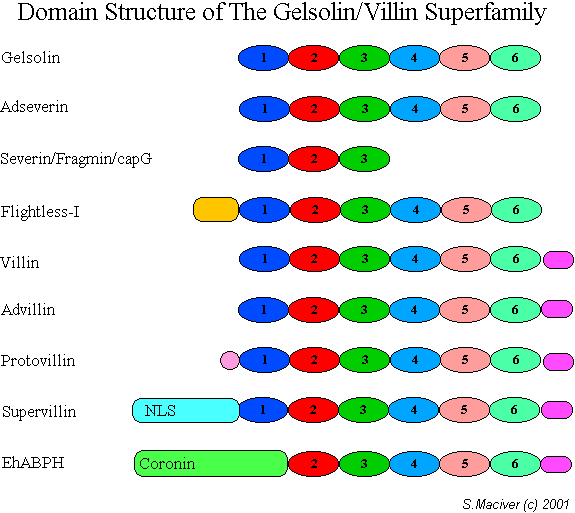
Gelsolin
(top) was the first member of the group to be sequenced (Kwiatkowski).
Its six domains are a repeat of two sets of three in that G1 (dark blue)
is most like G4 (light blue), G2 like G5, and G3, like G6 (Way
& Weeds,
1988).
The villin group are characterised by the possesion of the villin head
piece (Purple oblong) at the C-terminus. One of the most divergent forms
is EhABPH from Entamoeba histolytica (Ebert
et al, 2000)
which has a coronon-like N-terminal region followed by gelsolin/villin
domain but lacking G1.
For extra
information look under each particular ABP in the Encyclopaedia of
A.B.P.s.
|
| References:-
Ebert, F., Guillen, N., Leippe, M.
& Tannich, E. (2000) Molecular cloning and cellular localization of
an unusual bipartite Entamoeba histolytica polypeptide with
similarity to actin binding proteins., Mol.Biochem.Parasitol. 111,
459-464.
Kwiatkowski, D. J., Stossel, T. P.,
Orkin, S. H., Mole, J. E., Colten, H. R. & Yin, H. L. (1986) Plasma
and cytoplasmic gelsolins are encoded by a single gene and contain a
duplicated actin-binding domain, Nature. 323, 455-458.
Way, M. & Weeds, A. G. (1988)
Nucleotide sequence of pig plasma gelsolin. Comparison of protein
sequence with human gelsolin and other actin-severing proteins shows
strong homologies and evidence for large internal repeats, J. Mol.
Biol. 203, 1127-1133. |
