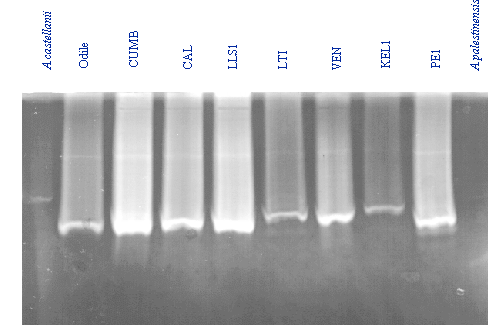
| The
so called Zymogram technique has been used by many workers to identify
proteases from amoeba (mainly Acanthamoeba where the extra
cellular protease is implicated in corneal degradation).We have modified this method for use in the
"Mini-Protean" gel system by BioRad. From
the methods of Heussen & Dowdle, 1980, Kleiner & Stetler-Stevenson
1994, Mitra et al (1994).
It is convenient to use Fish Skin Gelatin (Sigma G-7765).
1).
It is important that b-ME and DTT be kept out of any sample buffer for which protease
activity is to be detected. Samples
must therefore be made from non-reducing sample buffer:-
Working Concentration
Stock
100mls
20% Glycerol
-
20g
5% SDS
-
5g
0.4M Tris pH6.8
0.5M
80mls
(Bromephenol Blue added to give colour)
2). Make up gel mix including the gelatin at a suitable
concentration (0.1%). The
following is for
the BioRad “miniprotean” system and is enough for two
gels:-
Stock solutions
8%
10%
12%
14%
Distilled
Water
4.64mls 4.31mls
3.31mls
2.64mls
1.5M
Tris pH8.8
2.5mls
2.5mls
2.5mls
2.5mls
10%
SDS
100µl
100µl
100µl
100µl
45%
Fish skin gelatin
22µl
22µl
22µl
22µl
30%
Acrylamide mix
2.67mls
3.0mls
4.0mls
4.67mls
10%
APS
50µl
50µl
50µl
50µl
TEMED
20µl
20µl
20µl
20µl
Total
10mls
10mls
10mls
10mls
3). Run gel at 200v as usual, and then incubate the gel with 2.5%
Triton X-100 ; 50mM Tris-HCl pH 7.0
for 1 hour and then overnight at in 50mM Tris-HCl pH 7.0, 2mM
CaCl2 at room temperature.
Coomassie blue Staining
After
overnight incubation, stain with Coomassie as normal:-
0.1%
Coomassie R-250
40%
Methanol
10%
Acetic acid
Rock the gel in the stain for about one hour, then destain by briefly
washing the gel in tap water and then rocking in destain solution:-
10%
Acetic acid
5%
Methanol
|
| References:-
Alfieri, S.C.,
Correia, C.E.B., Motegi, S.A., & Pral, E.M.F. (2000). "Proteinase
activities in total extracts and in medium conditioned by Acanthamoeba
polyphaga trophozoites." J.Parasitol. 86,
220-227.
Buckwold, V. E., Alvarado, M., Carraso,
R. M. & Amils, R. (1999) A method for the determination of the pH
optima of proteases using unexposed photographic film., Anal.Biochem.
267, 420-421.
Cao, Z., Jefferson, D. M. &
Panjwani, N. (1998) Role of carbohydrate-mediated adherence in
cytopathogenic mechanisms of Acanthamoeba, J.Biol.Chem. 273,
15838-15845.
Cheung, A. L., Ying, P. &
Fischetti, V. A. (1991) A method to detect proteinase activity using
unprocessed X-ray films., Anal.Biochem. 193, 20-23.
He, Y.-G., Niederkorn, J. Y., McCulley,
J. P., Stewart, G. L., Meyer, D. R., Silvany, R. & Dougherty, J.
(1990) In vivo and in vitro collagenolytic activity of Acanthamoeba
castellanii, In.Oph.Vis.Sci. 31, 2235-2240.
Heussen, C. & Dowdle, E.B. (1980) Anal.Biochem.
102;196-202.
Hong, C.-Y.,
Kong, H.-H., Ock, M.-S., Kim, I.-S., & Chung, D.-I. (2001).
"Isolation and characterization of a cDNA encoding a subtilisin-like
serine proteinase (AhSUB) from Acanthamoeba healyi". Mol.Biochem.Parasitol.
111, 441-446.
Kleiner, D.E. & Stetler-Stevenson,
W.G. (1994) Anal.Biochem. 218;
325-329.
Mitro, K., Bhagavathiammai, A., Zhou,
O.-M., Bobbett, G., McKerrow, J. H., Chokshi, R., Chokshi, B. &
James, E. R. (1994) Partial characterization of the proteolytic
secretions of Acanthamoeba polyphaga, Exp.Parsitol. 78,
377-385.
Na, B.-K., Kim, J.-C. & Song,
C.-Y. (2001) Characterization and pathogenetic role of proteinase from Acanthamoeba
castellanii., Microbial Pathogenesis. 30, 39-48.
|